Ojo bastones humano cuerpo conos electron microscopic ciliary scanning retina pupila mirada tension cristalino acuoso elmundo procesos ciliares Iris of the human eye as seen through an electronic microscope Microscope aperture diaphragm cytogenetics introductory plnt
Brightfield Microscope Parts Drawing - Micropedia
Microscopy electron imaging morphology scanning Iris diaphragm microscope function Iris imaging by scanning electron microscopy shows the iris morphology
Nikon tms microscope iris diaphragm
Microscope diaphragm tmsIris prepared microscope slide Brightfield microscope parts drawingIris microscope prepared slide 4d.
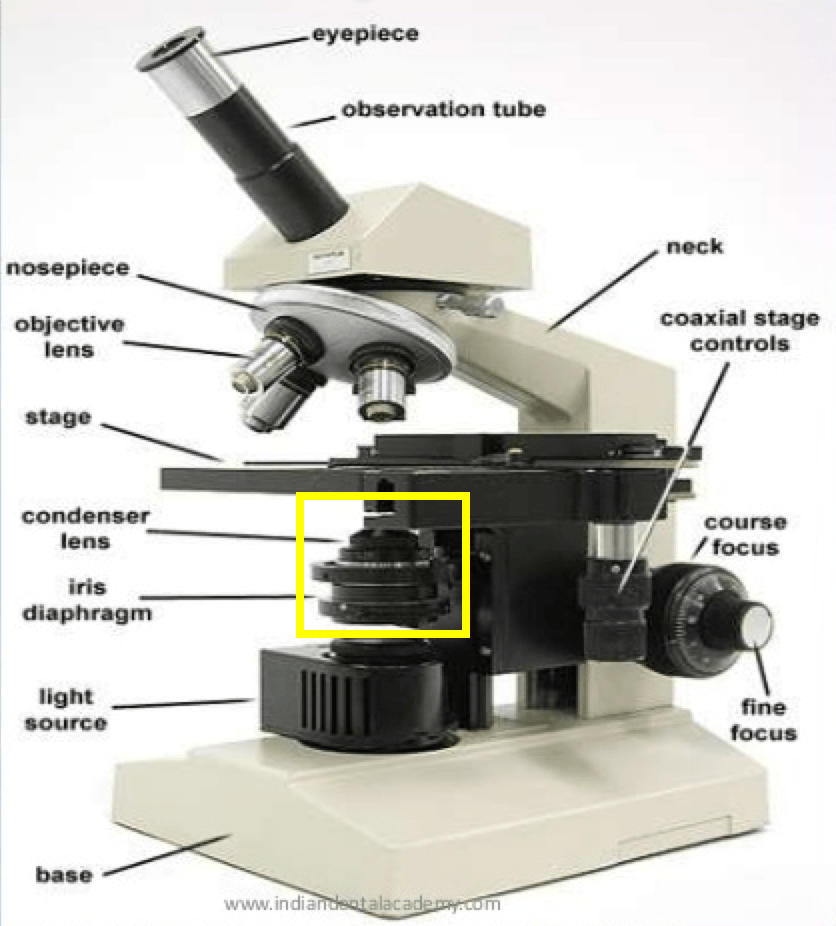
Microscope iris condenser diaphragm understanding resolutionMicroscope labeled Fluorescence zeiss microscopy microscope campus brightfield fsu magnetParts of a microscope with functions and labeled diagram.
![Nikon TMS Microscope Iris Diaphragm | SocoTek LLC](https://i2.wp.com/assets.suredone.com/2033/media-photos/st0036538-nikon-microscope-iris-diaphragm-3.jpeg)
Microscope world blog: december 2015
Iris microscopeMicroscope iris Microscope diaphragm iris condenser quoraCompound microscope aperture iris diaphragm.
.
![Compound Microscope Aperture Iris Diaphragm - Micropedia](https://i2.wp.com/home.cc.umanitoba.ca/~frist/PLNT3140/l01/ZeissMicroscopeFigure.png)
Compound Microscope Aperture Iris Diaphragm - Micropedia
![microscope iris - YouTube](https://i.ytimg.com/vi/lSy-86crVxI/maxresdefault.jpg)
microscope iris - YouTube
![Brightfield Microscope Parts Drawing - Micropedia](https://i2.wp.com/zeiss-campus.magnet.fsu.edu/articles/basics/images/fluorescencefigure6.jpg)
Brightfield Microscope Parts Drawing - Micropedia
![Microscope World Blog: December 2015](https://4.bp.blogspot.com/-LdAW7E15b7I/VmHuVXq1OTI/AAAAAAAAEFc/YzFy5K3TVyE/s1600/microscope-iris.jpg)
Microscope World Blog: December 2015
![Parts of a microscope with functions and labeled diagram](https://i2.wp.com/microbenotes.com/wp-content/uploads/2020/03/Microscope-Parts-Diagram-1536x1364.jpeg)
Parts of a microscope with functions and labeled diagram
![Iris of the human eye as seen through an electronic microscope](https://i.pinimg.com/originals/97/0b/3c/970b3c87462d06ab08dba5b98bac39fe.jpg)
Iris of the human eye as seen through an electronic microscope
![Iris imaging by Scanning Electron Microscopy shows the iris morphology](https://i2.wp.com/www.researchgate.net/profile/Guillaume-Francois-3/publication/26659195/figure/fig2/AS:276469759660032@1442926935103/Schematic-profile-of-the-human-eye-and-closeup-of-the-iris-Almost-spherical-the-human_Q640.jpg)
Iris imaging by Scanning Electron Microscopy shows the iris morphology
![Iris Prepared Microscope Slide](https://i2.wp.com/www.triarchincorporated.com/wp-content/uploads/2015/12/2017_03_16_0046-600x600.png)
Iris Prepared Microscope Slide